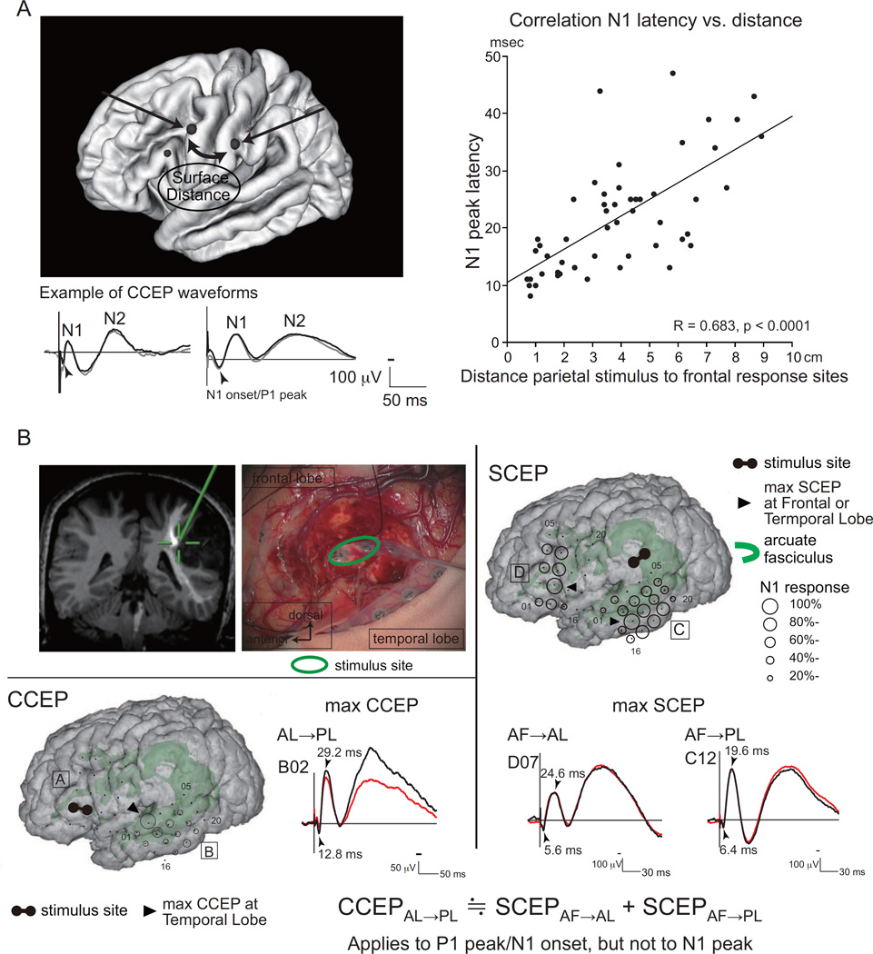
Figure 1. Evidence favoring direct cortico-cortical connections in generation of CCEPs.
A. Lateral parieto-frontal connectivity was explored by stimulating the parietal pairs and recording CCEPs from the frontal lobe. Representative CCEP waveforms (two subaverages each, N1 and N2 potentials) are shown (left, bottom). The vertical bar corresponds to the time of singe pulse electrical stimulation. Surface distance from the parietal stimulus sites (midpoint of the pair is used for calculation) to the maximum frontal CCEP response was measured. A positive correlation was observed between the surface distance and the N1 peak latency of the maximum response. When the regression line is extrapolated to the zero distance, there still remains ∼10 ms. This likely corresponds to the relatively long peak latency (∼10 ms) observed in the locally evoked DCR in humans [8]. Namely, direct cortical stimulation produces oligo- or multi- synaptic responses in the local cortical circuits. Adapted with permission from Ref. [30].
B. Subcortico-cortical evoked potentials (SCEPs) in a representative case. Left upper panel: Stimulation site (electrode pair, green circle) at the deep white matter at the floor of the tumor removal cavity. The site (green cross) was attached to the AF tract in the coregistered image on the neuro-navigation system.
Right panel: SPES (1 Hz, pulse width 0.3 ms, alternating polarity, 15 mA, 2 x 30 trials) was delivered to the stimulus site, and SCEPs were recorded both from the AL (SCEPAF→AL, D plate) and PL (SCEPAF→PL,C plate) at and around the terminations of the AF tract. The diameter of the circle at each electrode represented the percentile to the largest amplitude (N1) at the maximum SCEP response site.
Left lower panel: SPES of the AL (confirmed by 50 Hz electrical stimulation) produced CCEPs in the temporal lobe (presumed PL). The diameter of the circle at each electrode represented the percentile to the largest amplitude (N1) at the maximum CCEP response site. At the maximum response sites, the summation of P1 peak/N1 onset latencies of SCEPs (SCEPAF→AL + SCEPAF→PL, 12.0 ms) was very close to the P1 peak/N1 onset latency of CCEPAL→PL (12.8 ms). Adapted with permission from [26].
AF = arcuate fasciculus, AL = anterior language area, DCR = direct cortical response, PL = posterior language area, SPES = single pulse electrical stimulation

