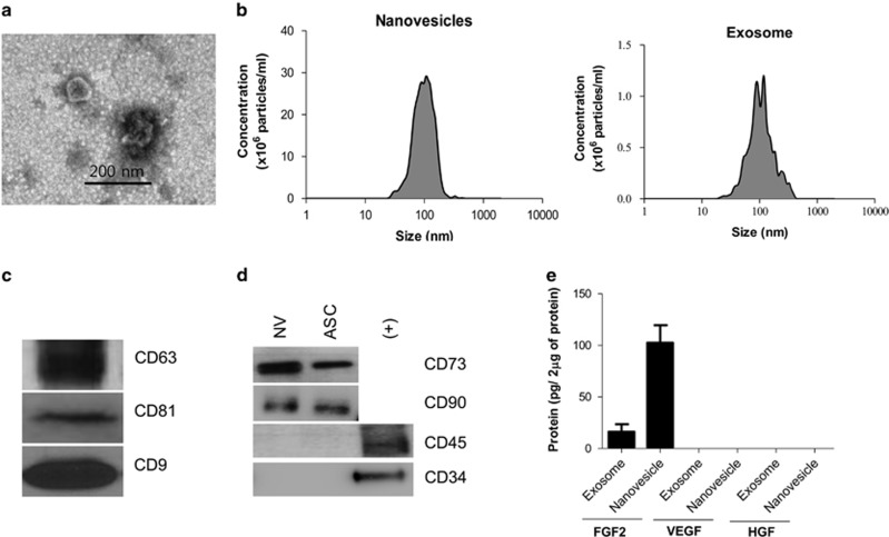
Figure 1.
Characterization of artificial nanovesicles generated from ASCs. (a) Transmission electron microscopy (TEM) image. (b) Nanoparticle size and number measured by nanoparticle tracking analysis. (c) Western blot analysis of artificial nanovesicles from ASCs using an exosomal marker. (d) Western blot analysis of artificial nanovesicles from ASCs using an ASC marker. NV: ASC-derived artificial nanovesicles, ASC: Adipose-derived stem cells. (e) ELISA measurements of the VEGF, FGF2 and HGF levels in ASC-derived natural exosomes and artificial nanovesicles.

