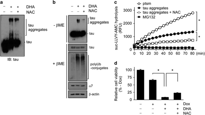
Figure 4.
Effects of docosahexaenoic acid (DHA) on tau aggregation and proteotoxic stress-mediated cytotoxicity. (a) Recombinant htau40 proteins (100 nM) were incubated with DHA (200 μM), N-acetylcysteine (NAC; 200 μM), or both. Tau aggregation was analyzed with SDS-PAGE/IB using tau antibodies under reducing conditions. (b) Monomeric and aggregated forms of tau proteins were observed in whole-cell extracts (WCEs) of inducible tau cells after doxycycline (Dox) treatment (500 ng ml−1) for 24 h. Cells were treated with DHA (200 μM), NAC (200 μM), or both for 4 h before sample collection. Protein samples were prepared under non-reducing conditions (without β-mercaptoethanol (-βME)) or reducing conditions (with β-mercaptoethanol (+βME)), and analyzed with SDS-PAGE/IB. (c) Proteasome activity was monitored with suc-LLVY-AMC hydrolysis after incubating htau40 (100 nM) with DHA (200 μM) in the presence or absence of NAC (200 μM). Values represent means±s.d. (n=3). *P<0.05 (n=3, two-way ANOVA). (d) DHA significantly sensitized the proteotoxicity of the inducible tau cell line, which was induced by excess amounts of overexpressed htau40. The inducible tau cell line was treated with 500 ng ml−1 Dox for 24 h in the presence or absence of DHA (50 μM) and NAC (50 μM). Cell viability was measured with the MTT assay. Values represent means±s.d. (n=3). *P<0.01 (one-way ANOVA with Bonferroni's multiple comparison test).

