
Figure 4.
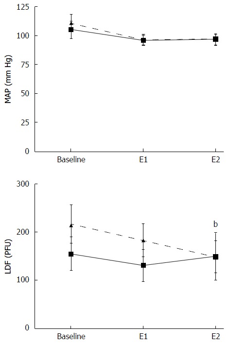
Gastric mucosal blood flow before and after oestrogen administration. Gastric mucosal blood flow [laser doppler flow (LDF)] presented as perfusion units (PFU) and mean arterial pressure (MAP) in male (dashed line) and female (solid line) animals. The X-axis shows values at baseline and following oestrogen administration at 0.1 µg/kg•min (E1) as well as 1 µg/kg•min (E2) respectively. Mean blood flow in the gastric mucosa decreased by 31% (68 ± 13 PFU) in males (n = 7) which was significantly different compared to baseline (b).