Figure 3.
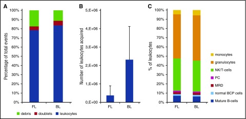
Evaluation of the EuroFlow bulk-lysis protocol. For reasons of comparison, each of the BM samples (day 15: n = 15; day 33: n = 15; day 78: n = 12) was processed according to the standard EuroFlow protocol (FL) and in parallel according to the EuroFlow bulk-lysis protocol (BL). (A) Number of leukocytes, debris, and doublets, calculated as percentage of acquired events. Using the bulk-lysis method, significantly less debris (P = .032 by paired Student t test) and significantly more leukocytes (P = .03 by paired Student t test) were measured. There were no significant differences between the 2 methods for the percentage of doublets. (B) Absolute number of leukocytes acquired. Using BL, on average 12-fold more leukocytes could be acquired (P < .0001). Please note that we included relatively many day 15 samples in order to be able to evaluate the impact of the 2 methods on the MRD levels as well. However, these day 15 samples generally have a very low white blood cell count; consequently, the number of leukocytes acquired after BL is still relatively low in a subset of samples. (C) Distribution of leukocyte subpopulations, defined as percentage of leukocytes. By paired Student t test (2-sided), small but statistically significant differences were observed for T/NK cells (mean: 24% vs 26%, P = .0047), granulocytes (mean: 33% vs 38%, P < .001), and monocytes (mean: 3.2% vs 4.5%, P < .001), whereas no significant differences were observed for the remaining populations. Of note, in 2 samples, MRD was only detected using the bulk-lysis method (0.013% and 0.018%) but not using the whole BM method. In the 11 samples MRD positive by both methods, MRD levels were not significantly different from each other (paired Student t test: P = .30), with mean values of 6.3% and 6.7% by whole BM and bulk-lysed BM method, respectively. Correlation analysis showed a Spearman r of 0.964 (95% confidence interval: 0.857-0.991; P < .0001). PC, plasma cells.

