Fig. 1.

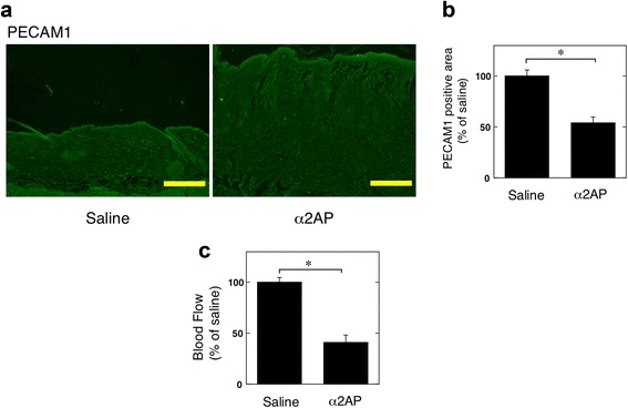
Effect of α2AP on vascular damage in mice. a The skin sections from saline or α2AP-administered mice were stained with antibodies to PECAM1. b The histogram shows quantitative representations of PECAM1 (n = 6). c Blood flow in the skin of saline or α2AP-administered mice (n = 4). The data represent the mean ± SEM. * P < 0.01. Scale bar, 200 μm
