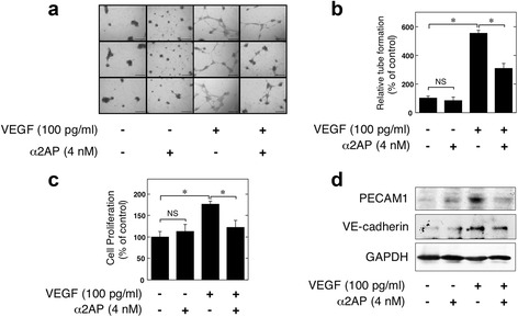
Fig. 4.

Effect of α2AP on the VEGF-induced pro-angiogenic effects in endothelial cells (ECs). a ECs were seeded on 96-well Matrigel-coated plates. ECs were cultured in the absence or presence of VEGF (100 pg/mL) or α2AP (4 nM) as indicated for 24 hours. b Tube formation in ECs was measured as described in Materials and Methods (n = 6). c ECs were cultured for 24 hours in the absence or presence of VEGF (100 pg/mL) or α2AP (4 nM). Cell proliferation was assessed as described in Materials and Methods (n = 3). d ECs were cultured for 24 hours in the absence or presence of VEGF (100 pg/mL) or α2AP (4 nM). The expression of each protein was examined by western blot analysis. The data represent the mean ± SEM. * P < 0.01. NS, not significant. Scale bar, 50 μm
