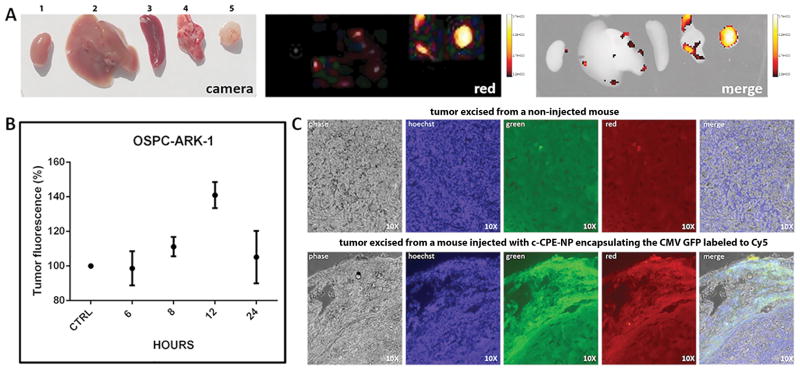
Figure 5. c-CPE-NP transfection efficiency in vivo in ovarian tumor cells.
SCID mice harboring subcutaneous OSPC ARK-1 xenografts were injected intraperitoneally with 2.5mg of c-CPE-NP encapsulating the CMV GFP plasmid labeled to the Cy5 dye. Mice were sacrificed at different time points and organs including, kidney, liver, spleen, lungs and tumors (labeled from 1 to 5, respectively) were excised for histological examination and visualization using an In-Vivo FX-PRO imaging system (excitation/emission:550/635nm; exposure time 10 seconds). A and B, Tumors fluorescence peaks at 12 hours after particles’ injection and is significantly higher than fluorescence detected in healthy organs or tumors excised from control (ie, non-injected) mice (panel B; p=0.007). (C) Fluorescence microscopy images on tumor slides excised from mice 12 hours after treatment with c-CPE-NP encapsulating the CMV GFP plasmid labeled with the Cy5 dye. Most of the cells in which the DNA was successfully delivered by the c-CPE-NP (lower panel, red image) also expressed GFP (lower panel, green and merge images). Tumors excised from non-injected control mice were visualized using the same parameters to evaluate tissue autofluorescence (upper panel).

