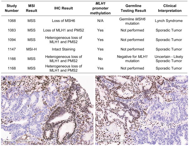
Figure 1.
The table summarizes endometrial cancer patients with discordances between MSI and IHC tissue testing results. The tumor in the photomicrograph demonstrates heterogeneous loss of MLH1 (A) and PMS2 (B) protein expression by immunohistochemistry. Retained protein expression is indicated by red-brown nuclear staining; loss of protein expression is apparent in tumor cells with blue nuclei. Loss of PMS2 protein expression is secondary to the primary defect in MLH1, as MLH1 and PMS2 typically exist as dimers in the nucleus.

