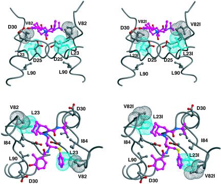
FIG. 1.
Packing of residue 23 within the crystal structure of HIV-1 protease bound to nelfinavir (1OHR) (6). Shown are two views of the active site separated by 90°. The images on the left are the crystal structure, and those on the right are with side chains of L23 and V82, each replaced with isoleucine within the graphics program MIDAS (4). Residue 23 is colored cyan; the other atoms are colored by atom type, with the carbons in nelfinavir colored magenta. van der Waals surfaces are shown for the side chains of residues 23 and 82.

