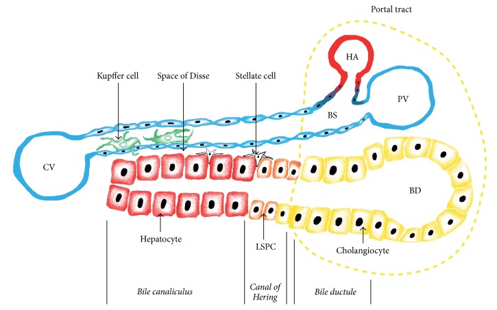
Figure 1.
Schematic histological structure of liver tissue. Functional units of liver tissue are formed by trabeculae and accompanying blood sinusoids. Liver tissue gets its afferent blood supply from two sources: hepatic artery and portal vein. Hepatic arterioles (HAs) and the terminal branches of portal vein (PV) merge to form blood sinusoids (BSs) lined with endotheliocytes and drained into the central veins (CVs). In the sinusoids, close to endothelium reside liver macrophages named Kupffer cells. Bile produced by hepatocytes flows in the opposite direction and is discharged into the bile ducts (BDs). Hepatic arterioles, terminal branches of portal vein, and the smallest bile ducts are drawn together forming compact structures called portal tracts shown at the right side of the figure. Liver trabeculae are built of hepatocytes. The inner cavities of trabeculae form canaliculi which are closed at the central ends of the lobules (left side of figure) and while on their way to BD they convert into bile ductules (BDLs) via a transitory zone called the canals of Hering (CH). Bile ductules drained into the bile ducts are lined with cholangiocytes, and the canals of Hering contain LSPCs. Tiny spaces between trabeculae and the endothelium of blood sinusoids are called the spaces of Disse (SD). They participate in the bidirectional traffic of different substances between blood and hepatocytes and contain stellate (Ito) cells.

