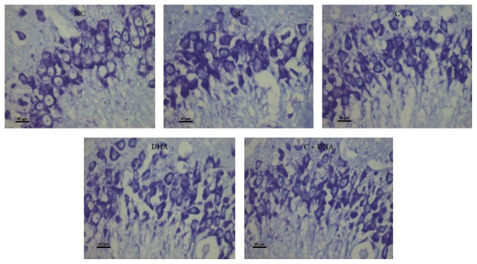
Figure 4.
CA3 region: representative photomicrographs of CA3 region from 5 μ thick hippocampal sections viewed by 40x magnification, showing Cresyl Violet stained neurons from all experimental groups of PND 40 rats. Note: rats supplemented with combined choline and DHA show significant increase in the number of neural cells in CA3 region.

