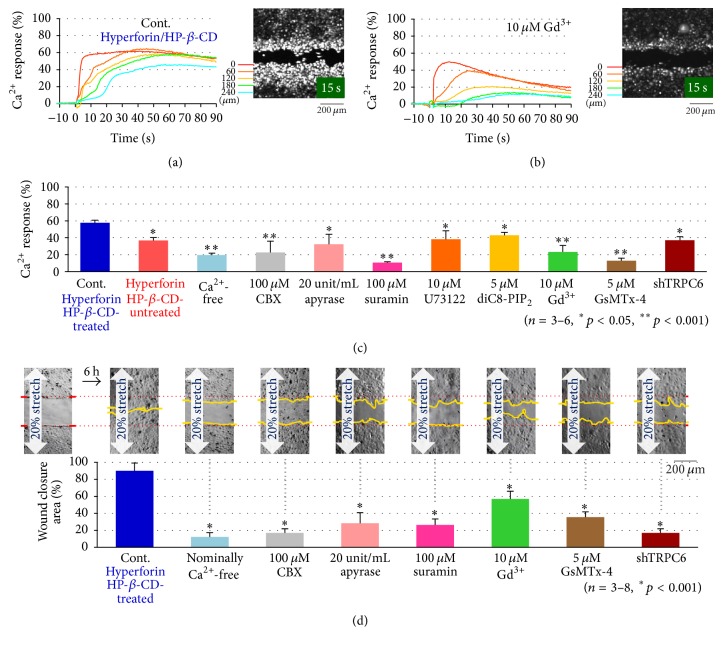
Figure 3.
The effects of various inhibitors on the stretch-induced Ca2+ responses and wound closure. (a) The time course of changes in the fluorescence intensity of Fluo-8 due to a transient 20% stretch in hyperforin/HP-β-CD-treated HaCaT cells (control). Each color trace indicated the data at different distances of 0, 60, 120, 180, and 240 from the wound edge (inset image). The intensity was normalized to the peak value obtained with ionomycin treatment at the end of each experiment. (b) The effects of Gd3+ on the stretch-induced Ca2+ response as a typical example of the blocking effects of the inhibitors. Gd3+ (10 μM) was applied at 10 min before the application of a 20% stretch. (c) The effects of various inhibitors on 20% transient stretch-induced Ca2+ responses in hyperforin/HP-β-CD-treated HaCaT cells. The intensity traces at each distance from the wound edge were averaged and normalized to the peak intensity obtained with ionomycin treatment. The data show the average of the peak values obtained in 3–6 separate experiments. Various inhibitors, including CBX (100 μM), apyrase (20 Unit/mL), suramin (100 μM), U73122 (10 μM), diC8-PIP2 (5 μM), Gd3+ (10 μM), and GsMTx-4 (5 μM), were applied at 10 min before the stretch stimulation. A Ca2+-free condition was achieved by changing the medium to Ca2+-free medium that contained 0.5 μM EGTA. All of the quantitative data are shown as the mean (±SEM). (d) The effects of various inhibitors on the stretch facilitated wound closure in hyperforin/HP-β-CD-treated HaCaT cells. Confluent cell cultures were scratched and allowed to migrate for 6 h under a sustained 20% stretch in a medium that contained various inhibitors, including CBX (100 μM), apyrase (20 Unit/mL), suramin (100 μM), Gd3+ (10 μM), and GsMTx-4 (5 μM) or in nominally Ca2+-free medium. shTRPC6 was applied to the cells for 3 h; the cells were then grown to confluence. Representative DIC images (upper panel) and the means of 3–8 wound closure experiments at 6 h after scratching (lower panel) are shown. All of the quantitative data are shown as the mean (±SEM).

