
Fig. 1.
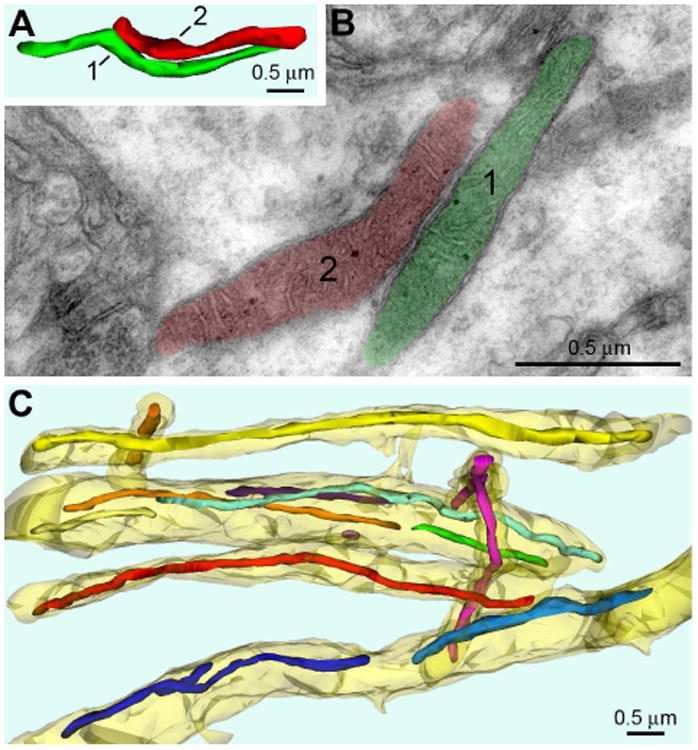
Shape and ultrastructure of mitochondria in dlPFC layer III dendritic shafts from 11 yo rhesus macaque. (A, B) 3D image and electron micrograph of two representative mitochondria (correspondingly labeled “1” and “2” and depicted green and red) located in the same dendrite. Shape of the mitochondria and ultrastructure of membranes and matrix are characteristic for normally functioning neurons. (C) 3D reconstruction from 33 serial sections from a randomly chosen segment of neuropil reveals numerous mitochondria elongated along dendritic shafts (shown semitransparent yellow). Notice similar diameters of all the mitochondria throughout entire length. Each mitochondrion is depicted with different color facilitating identification in 3D image.