
Fig. 5.
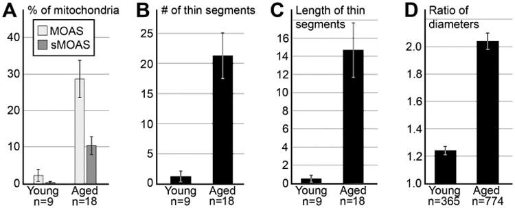
Mitochondrial morphological parameters in dendrites from dlPFC of young adult (7 and 11 yo) and aged (26, 27, 31 and 33 yo) rhesus macaques. (A) Percentages of MOAS and sMOAS dramatically differ between young and aged animals. (B, C) Number and length of thin segments in MOAS normalized per 100 micron length of reconstructed mitochondria. (D) Ratio of maximal vs. minimal diameters of mitochondria. Error bars show SEM. n is number of neuropils segments (A-C) or number of mitochondria analyzed (D).