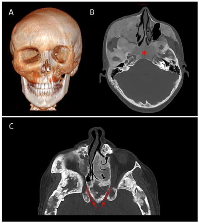
Figure 4.

Craniofacial polyostotic fibrous dysplasia.
Imaging from two individuals with polyostotic craniofacial fibrous dysplasia (FD) in the setting of McCune-Albright Syndrome.
(A) 3D CT of a 17 year-old male, showing extensive disease of the midface and orbital asymmetry.
(B) Axial CT scan showing typical ground-glass appearance of the left midface and skull base, obliterating the region of the sphenoid sinuses (asterisk).
(C) Axial CT from a 33 year-old female showing the narrowing of the bilateral optic canals (outlined in red).
