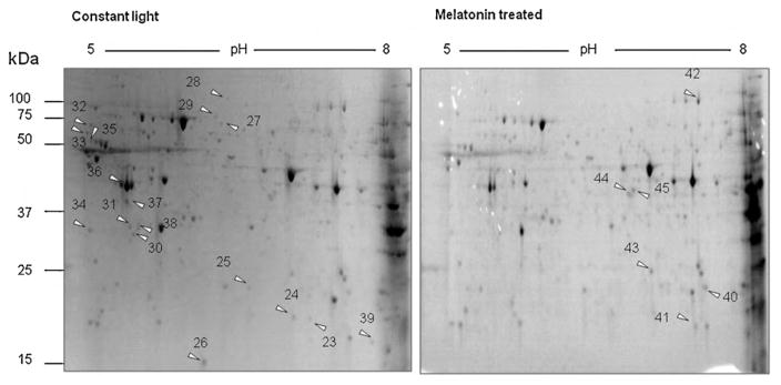
Fig. 3.
2D-SDS-PAGE analysis of retinal proteins from constant light group and melatonin treated group. Retinas were isolated from mice that were housed in constant light conditions treated with or without melatonin. Arrows are showing spots of up- or down-regulation. (A) Retinal proteome of mice exposed to constant light and treated with control vehicle; (B) retinal proteome of mice exposed to constant light and treated with melatonin.

