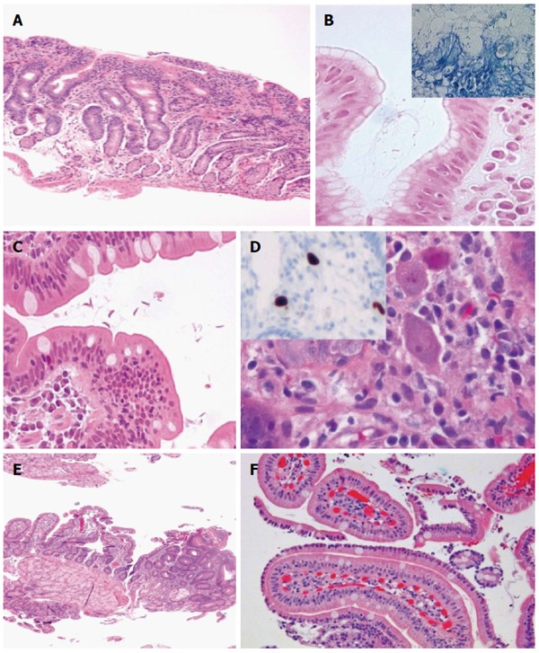
Figure 4.

Gluten-sensitive enteropathy mimickers. A: Lymphocytic gastritis with involvement of the duodenum (HE, × 100); B: H. pylori gastritis (inset, Giemsa staining) (HE and Giemsa × 630); C: Giardiasis (HE, × 400); D: Cytomegalovirus (CMV) infection (HE, × 6300) and inset showing anti-CMV antibody reacting against viral proteins using an avidin-biotin complex immunoperoxidase immunohistochemical detection (× 100); E: Focal adenomatous change in duodenum (HE, × 50); F: Sickle cell disease-related duodenitis (HE, × 200).
