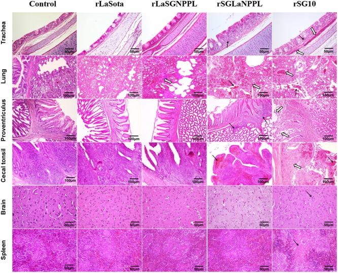
FIGURE 5.

Tissue histopathology of inoculated 4-week-old chickens. Chickens were infected oculonasally with parental rSG10, parental rLaSota or their chimeric viruses (rLaSGNPPL and rSGLaNPPL). Birds were sacrificed at 4 dpi and tissue was fixed with formalin, sectioned and stained with hematoxylin and eosin. The trachea had lymphocytic infiltration of the lamina propria (black arrows) and submucosal congestion (empty arrows) of the tracheal mucosa. The lung had hemorrhage, RBC infiltration in both the lung and bronchi (black arrows) and congestion (empty arrows). The proventriculus had infiltration of lymphocytes in the mucous membranes of the nipple and the glands (black arrows) and severe mucosal epithelial shedding (empty arrows). The cecal tonsils indicated congestion of the lamina propria (black arrows) and necrosis and shedding of lymphocytic cells of the mucosa (empty arrows). The brain indicated increased and gathered microglial cells (black arrows). The spleen had necrosis and focal cellular necrosis formation (black arrows).
