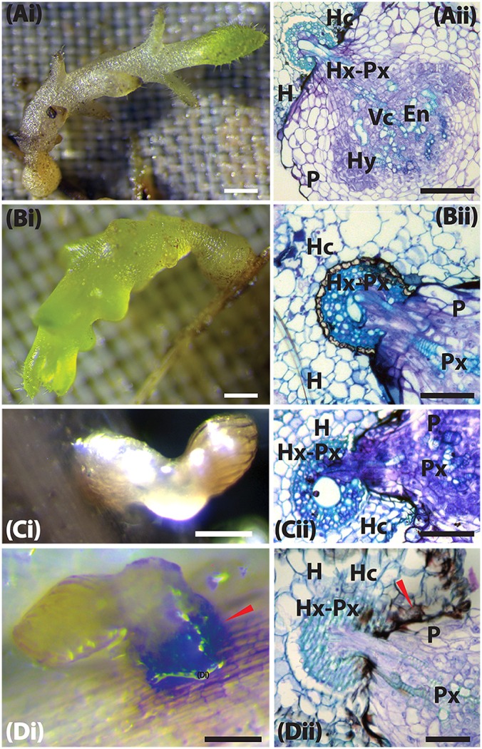
Figure 5.

Host resistance mechanism of sorghum to Striga hermonthica 9 DAI. (Ai) Colonization of WSD-1 sorghum root by S. hermonthica (Kibos ecotype) 3 days after infection showing a well-established haustorium (the scale bar is 1 mm). (Aii) Transverse section of an embedded root tissue of WSD-1 sorghum accession showing penetration of the host root cortex and endodermis and vascular connections between the host and parasite xylem (Hx-Px). Scale bar is 0.1 mm. (Bi) Genotype N13 9 days after infection. The haustorium is well-developed and shows swelling at the point of infection. The scale bar is 1 mm. (Bii) Transverse section of embedded tissue of N13 9 DAI. By this time, the parasite had a well-developed endophyte (En) and hyaline body (Hy). The dark areas around the pericycle may indicate a resistance mechanism. The scale bar is 0.1 mm. (Ci) A resistant wild sorghum accession (WSA-1) infected with S. hermonthica. The parasite was weak and did not form much vegetative tissue. The scale bar is 5 mm. (Cii) A transverse section of a resistant wild sorghum accession (WSA-1). Only a small part of the parasite was able to penetrate the host vascular system. The scale bar is 0.1 mm. (Di) Colonization of WSE-1 9 DAI showing more intensive secondary metabolite deposits (red arrow). In most cases, the parasite died and did not persist past 14 DAI. The scale bar is 5 mm. (Dii) A transverse section through the haustorium of the resistant wild sorghum accession WSE-1. Only a small part of the parasite xylem was able to make vascular connections with the host. In addition, the hyaline body (Hy) as well as the endophyte (En) were much smaller compared to N13. The scale bar is 0.1 mm.
