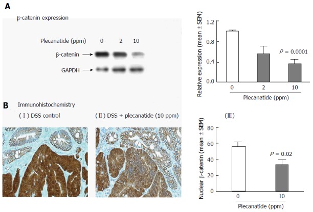
Figure 5.

Total β-catenin expression is reduced within the colon of Apc+/Min-FCCC mice treated with dextran sodium sulfate plus plecanatide. A: Western blot analysis and the associated densitometric quantification of levels of total β-catenin expression (mean ± SEM) within the colon; B: Immunohistochemical localization of β-catenin within the colonic mucosa. Membranous localization of β-catenin was observed within the normal colonic mucosa irrespective of the treatment group, while cytoplasmic and nuclear β-catenin staining predominant in adenomas from DSS-treated mice (panel I). Plecanatide treatment caused a significant reduction in nuclear staining of β-catenin in dysplasias, while the cell membranes exhibited enhanced protein localization (panel II). The number of tumor cells with nuclear localization of β-catenin was counted in distal colon tumors (n = 7-9 mice/group) and expressed as a percentage of the total number of tumor cells per 400 X field (panel III). Statistical comparisons between DSS control and DSS plus plecanatide-treated groups were performed using the Student’s t test. A P value of ≤ 0.05 was considered significant. DSS: Dextran sodium sulfate; GAPDH: Glyceraldehyde 3-phosphate dehydrogenase; .
