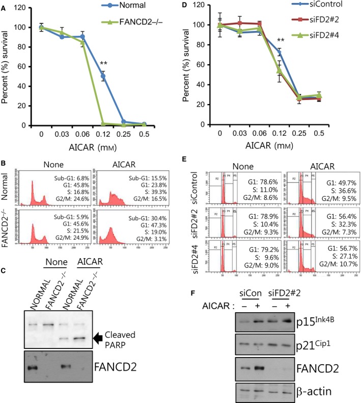
Figure 4.

AICAR‐induced apoptosis and cell cycle arrest are affected by FANCD2 repression. (A) Effects of FANCD2 knockout on cell proliferation/survival after AICAR treatment in transformed fibroblasts. MTT assay was performed for normal and FANCD2−/− fibroblasts treated with the indicated concentrations of AICAR for 3 days. The percent (%) survival was calculated for comparison with untreated cells. A representative graph from three independent experiments is shown. The values represent the mean ± SD (Student's t‐test, **P < 0.01). (B) Cell cycle analysis of normal and FANCD2−/− fibroblasts treated with AICAR. Cells were treated with 0.25 mm AICAR for 2 days, and flow cytometric analysis was performed after propidium iodide staining. A representative data from three independent experiments are shown. The percentages of cells in the sub‐G1 population are shown inside each graph. (C) Effects of FANCD2 loss on AICAR‐induced PARP cleavage. Transformed fibroblasts were treated with 0.25 mm AICAR for 2 days, and cleaved PARP was detected via immunoblotting. (D) Effects of FANCD2 knockdown on cell proliferation/survival after AICAR treatment in Caki‐1 cells. Caki‐1 cells were transfected with control siRNA (siControl) or FANCD2‐targeting siRNAs (siFD2#2 or siFD2#4), and MTT assay was performed. The percentage survival was calculated for comparison with untreated cells. A representative graph from three independent experiments is shown. The values represent the mean ± SD (Student's t‐test, **P < 0.01). (E) Effects of FANCD2 knockdown on cell cycle distribution of AICAR‐treated Caki‐1 cells. Cells were transfected with siControl, siFD2#2, or siFD2#4. Cell cycle analysis was performed as in (B). The percentages of cells at G1 and S phases are shown inside each graph. (F) Effects of FANCD2 knockdown on levels of cell cycle regulators. Caki‐1 cells were transfected with control siRNA (siCon) or FANCD2 siRNA (siFD2#2) and treated with AICAR for 24 h. The levels of p15Ink4B and p21Cip1 were evaluated by immunoblotting.
