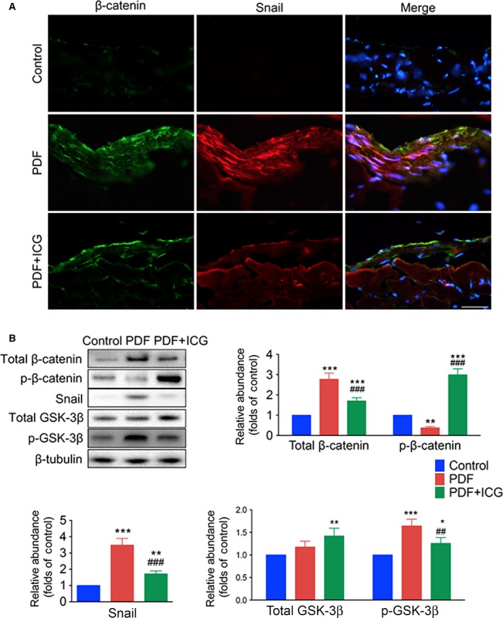
Figure 3.

ICG‐001 reactivates GSK‐3β and decreases the expression of Snail and β‐catenin in the peritoneum after PDF exposure. (A) Images of frozen sections stained with antibodies against β‐catenin (green) and Snail (red), nuclei stained with Hoechst (blue) (original magnification, ×400; scale bar, 100 μm). (B) Western blots of peritoneal tissue lysates with antibodies against phosphorylated β‐catenin (p‐β‐catenin), total β‐catenin, Snail, phosphorylated GSK‐3β (p‐GSK‐3β), total GSK‐3β, and β‐tubulin. Statistics of expression levels of p‐β‐catenin, total β‐catenin, Snail, p‐GSK‐3β, and total GSK‐3β normalized against β‐tubulin (mean ± SD, n = 4, ***P < 0.001, **P < 0.01, and *P < 0.05 vs control group, ### P < 0.001 and ## P < 0.01 vs PDF group).
