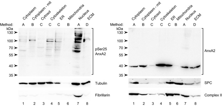
Figure 1.

Detection of pSer25AnxA2 in subcellular fractions derived from PC12 cells. Proteins (100 μg) from the cytoplasm (lane 1), the cytoplasm devoid of mitochondria (‐mit; lane 2), the cytosol (lane 3), the cytoskeleton (lane 4), ER (lane 5), mitochondria (lane 6), the nuclear fraction (lane 7) and EGTA‐released ECM (lane 8) were separated by 10% SDS/PAGE and subjected to western blot analysis. The blots were probed with antibodies against pSer25AnxA2 and total AnxA2, as indicated. Antibodies against compartmental markers, namely the cytoplasm (tubulin; 55 kDa), ER (SPC; 25 kDa), nucleus (fibrillarin; 35 kDa) and mitochondria (complex II; 70 kDa) were also employed as indicated. The blot probed against pSer25AnxA2 was reprobed against fibrillarin, while the blot probed against AnxA2 was reprobed against SPC. Only 25 μg of protein from the mitochondrial fraction was used for western blot analysis of tubulin and complex II on two different membranes. Detection of the immunoreactive protein bands was performed using the ChemiDoc™ XRS+ molecular imager after incubation with HRP‐conjugated secondary antibodies and enhanced chemiluminescence (ECL) reagent. The methods (A–D) used to generate the different fractions are indicated above the western blots and described in the Methods section. The arrowheads to the left indicate the protein molecular mass standards.
