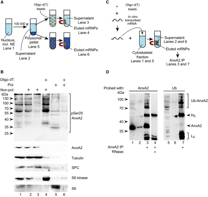
Figure 4.

pSer25AnxA2 and ubiquitinated high‐molecular‐mass forms of AnxA2 associate with translationally inactive mRNP complexes. (A, B) High‐molecular‐mass forms of pSer25AnxA2 is present in oligo(dT)‐purified nonpolysomal mRNP complexes in PC12 cells. (A) Schematic representation of the method used in (B) with reference to the individual lanes in (B). (B) Samples (100 μg of protein) were prepared from the following fractions: nucleus (lane 1), supernatant (lane 2) and polysome‐containing pellet (lane 5; derived from the nuclear fraction after centrifugation for 2 h 100 000 g through a 1 m sucrose cushion), non‐oligo(dT)‐bound supernatant (lane 3), oligo(dT)‐bound supernatant (lane 4), and oligo(dT)‐bound pellet (lane 6), as indicated above the western blot. The proteins were separated by 10% SDS/PAGE and subjected to western blot analysis. The blots were probed with antibodies against pSer25AnxA2, total AnxA2, tubulin, SPC, S6 kinase and the ribosomal protein S6, as indicated. Detection of the resulting protein bands was performed by the ChemiDoc™ XRS+ molecular imager after incubation with HRP‐conjugated secondary antibodies and ECL reagent. The arrowheads to the left indicate the protein molecular mass standards. (C, D) High‐molecular‐mass forms of AnxA2 in mRNP complexes affinity‐purified via binding to anxA2 mRNA represent ubiquitinated forms of the protein. (C) Schematic overview of the method used in (D) with reference to the individual lanes in (D). Proteins (100 μg) present in the total cytoskeletal fraction derived from NGF‐stimulated PC12 cells (lanes 1 and 5), the unbound fraction (lanes 2 and 6) and AnxA2 IP proteins from the affinity‐purified mRNP complexes derived from the cytoskeletal fraction (lanes 3 and 7), were subjected to 10% SDS/PAGE and immunoblot analysis using monoclonal antibodies against AnxA2 (lanes 1–4) or Ub (lane 5–7). The bands representing ubiquitinated AnxA2 are indicated by the upper bracket to the right. Lane 4 represents a negative control, showing the binding of cytoskeleton‐associated proteins to anxA2 mRNA coupled to oligo(dT) magnetic beads in the presence of RNase, followed by IP using monoclonal AnxA2 antibodies. The molecular mass markers are indicated to the left and the IgG heavy (Hc; arrowhead) and light (Lc; lower bracket) chains to the right.
