Figure 5.

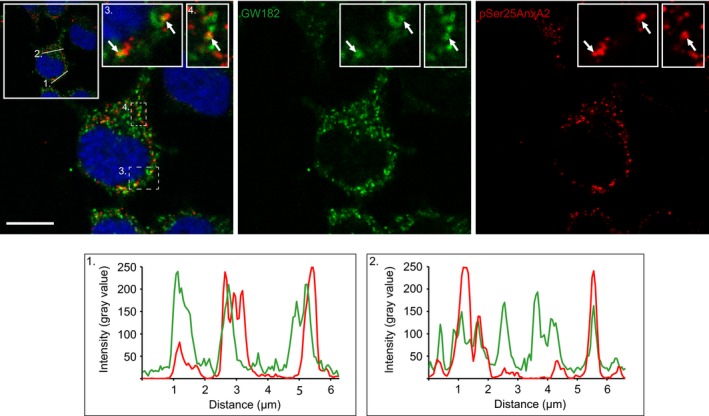
pSer25AnxA2 partially colocalises with the P‐body marker GW182. PC12 cells were double‐stained for immunofluorescence using mono‐ and polyclonal antibodies against GW182 (green) and pSer25AnxA2 (red) respectively. The insets in the merged confocal image to the left – including DAPI staining (blue) to highlight the nuclei – show higher magnifications of the regions, denoted in 3 and 4, to illustrate the partial colocalisation of pSer25AnxA2 and GW182. Scale bar: 10 μm. The fluorescence intensity profiles (from left to right) of the two proteins correspond to the cross‐sections, denoted 1 and 2, shown in the insert in the upper right corner of the merged image.
