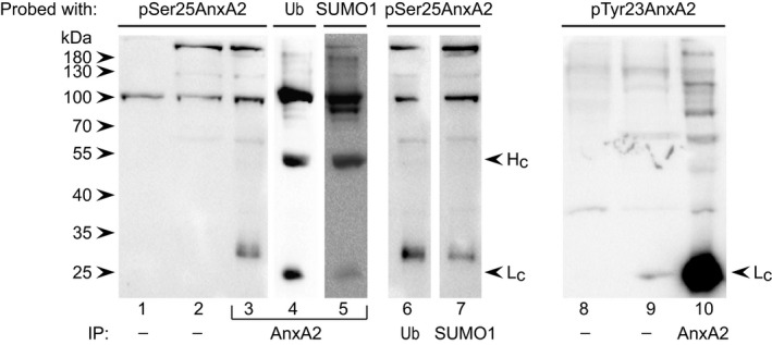
Figure 6.

pSer25AnxA2 is ubiquitinated and sumoylated, but shows a different high‐molecular‐mass pattern than pTyr23AnxA2. Proteins (100 μg) in the cytoplasmic (lanes 1 and 8) and nuclear (lanes 2 and 9) fractions (1/6 input) as well as IPs of AnxA2 (lanes 3–5 and 10), Ub (lane 6) or SUMO1 (lane 7) from the nuclear fraction of PC12 cells were subjected to 10% SDS/PAGE and western blot analysis using antibodies against pSer25AnxA2 or pTyr23Anxa2, as indicated. Note that the secondary anti‐mouse HRP‐conjugated antibody obtained from Jackson Immuno‐Research (205‐032‐176) is light chain specific and also note that the blots probed with the polyclonal anti‐pSer25AnxA2 shows no light chain as the antibodies against AnxA2, Ub and SUMO1 are all mouse monoclonal antibodies. The immunoreactive protein bands on the membrane were visualised using ECL reagents.
