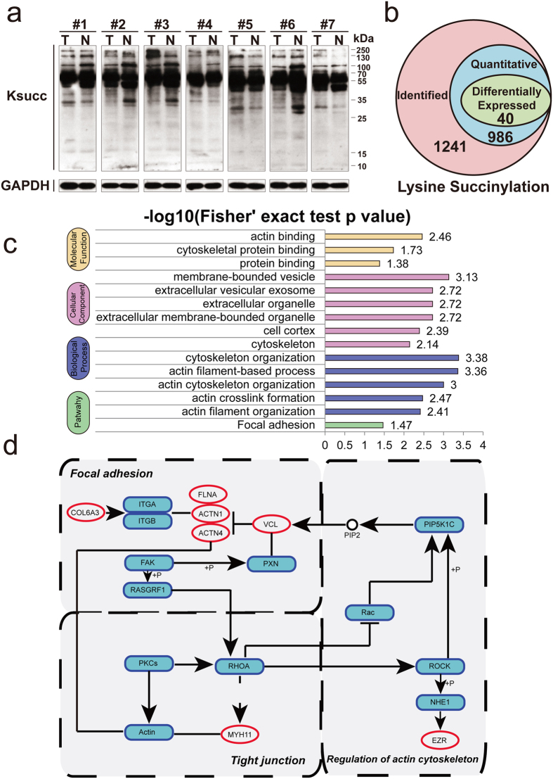
Figure 5. Identification and functional analysis of the lysine succinylome in gastric cancer.
(a) The expression profile of lysine succinylation in GC tissues by western blotting using a pan anti-succinyl lysine polyclonal antibody (full-length and multiple exposures gels are presented in Supplementary Fig. 4). (b) Summary of lysine succinylated sites in GC. (c) GO enrichment analysis of down-regulated succinylated proteins. (d) Three major pathways (focal adhesion, regulation of actin cytoskeleton and tight junction) of succinylated proteins. Red oval represents lysine succinylated proteins.

