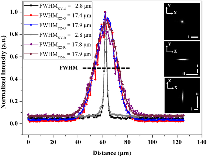
Figure 2. Lateral and axial resolution at the thinnest light-sheet region under the light-sheet microscope, with the 6.3X zoom-in.

Values of PSF in glycerol and RIMS were shown as FWHM. R: RIMS; G: glycerol. PSFs on the XY-, YZ- and XZ-planes were shown with the scale bar (i–ii) 25 μm.
