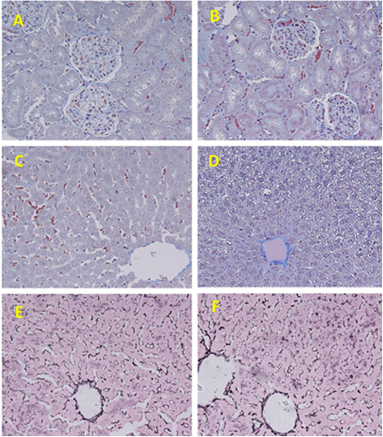
Figure 5. Representative photomicrograph of the histopathological examinations of both kidney and liver.
(A,B) Control and treated renal tissues stained with Masson Trichrome (X 400). (C,D) Control and treated hepatic tissues stained with Masson Trichrome (X 400). (E,F) Control and treated hepatic tissues stained with Reticulin (X 400).

