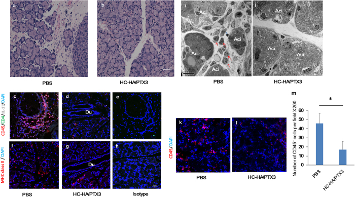
Figure 2. HC-HA/PTX3 reduces infiltration of inflammatory and immune cells into lacrimal glands of cGVHD mice.
Lacrimal glands collected at Day 32 after BMT, i.e., 4 days after the last injection of PBS or HC-HA/PTX3, were sectioned for H & E staining (a,b), scale bar = 50 μm), immunofluorescence staining against CD45 (Red), CD4 (Green), IL-17 (White) (c,d), MHC class II (Red) (f,g), and DAPI nuclear staining (Blue) (c–h), scale bar = 20 μm), and transmission electron microscopy (i and j), Ac: acinus; red arrows: infiltrating inflammatory cells; asterisks: capillary; scale bar = 10 μm). Immunostaining for CD45 (red) and cell nuclei (blue) (k and l), scale bar = 20 μm). The number of CD45+ cells per field at x200 magnification in the HC-HA/PTX3- and PBS-treated groups were counted at least 5 different field on 4 sections (m), **p = 0.015, N = 4 for each group).

