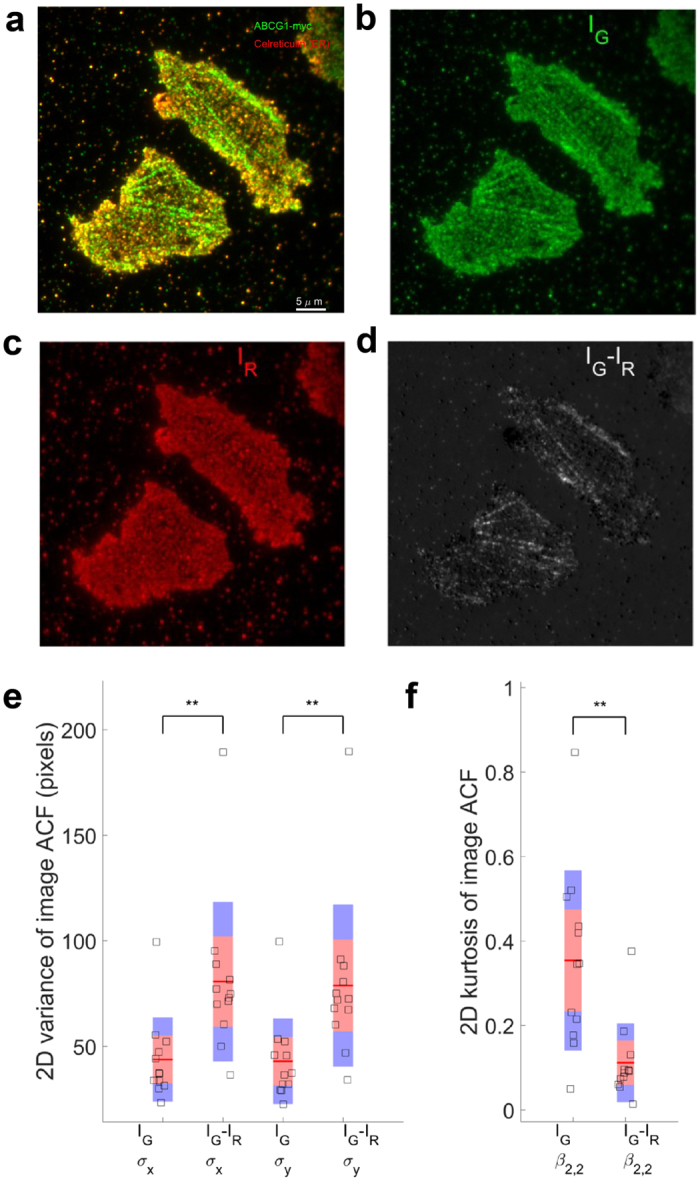
Figure 2. Filamentous ABCG1 does not localize with calreticulin in the ER.

(a) Representative example of intact, untreated, permeabilized and fixed cells immunostained for ABCG1-myc (green) and the ER marker calreticulin (red). (b) Green (IG) and (c) red (IR) channels images and (d) the difference (IG-IR) image. (e) The variances of the ACF of the difference images (n = 12) are larger than that of green channel alone, indicating that ABCG1 that did not co-localize with ER tended to be filamentous in nature. (f) 2D Kurtosis displays lower peakedness in the difference image than in the green channel alone **P < 0.01.
