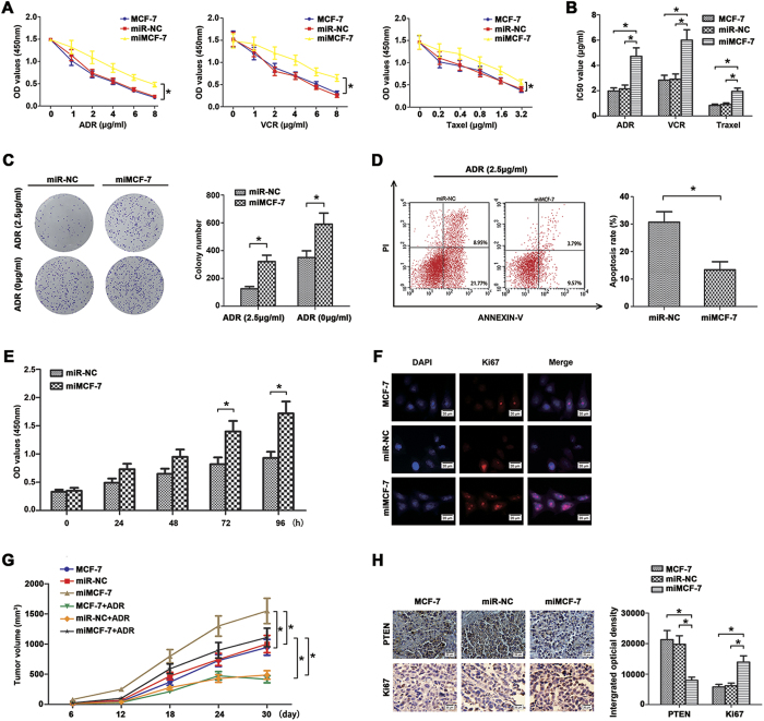
Figure 3. Over-expression miR-130b enhanced chemoresistance and proliferation of MCF-7 cells in vitro and in vivo.
(A) After transfection, cells were treated with different concentrations of ADR, VCR and Taxel for 48 h, respectively. Cell absorbances (450 nm) were assessed by CCK8 assay. (B) The reported values were the IC50 (mean ± SD) of three independent experiments. IC50 represents the drug concentration producing 50% decrease of cell growth. There were no significant differences between the control group and the blank group. (*P < 0.05; miR-130b mimics vs miR-NC). (C) Effects of miR-130b on the formation of cell clones when cells were treated with or without ADR (2.5 μg/ml) were analyzed by clone formation assay. Transfection of the MCF-7 cells with miR-130b mimics decreased their sensitivity to ADR treatment and enhanced their clone formation ability. (D) Decreased apoptosis rate were measured by flow cytometry assay. (E) The viabilities of MCF-7 cells transfected with miR-130b mimics or miR-NC were detected by CCK8 assay at 0, 24, 48, 72 and 96 h. (F) Ki67 expression was detected by immunofluorescence staining in these cells. Red fluorescence: Ki67, blue fluorescence: DAPI, staining for nuclear DNA. There were no significant differences between the control group and the blank group. (G) The miR-130b enhanced the tumourigenicity in the nude mice xenograft model. The tumor growth curves at 30 days are shown as indicated. (H) Deregulation of PTEN (×400) and Ki67 (×400) expressions were showed using IHC staining in xenograft tumors derived from MCF-7, miR-NC and miMCF-7 cells. There were no significant differences between the control group and the blank group (*P < 0.05). The results were showed as mean ± SD of three independent experiments.

