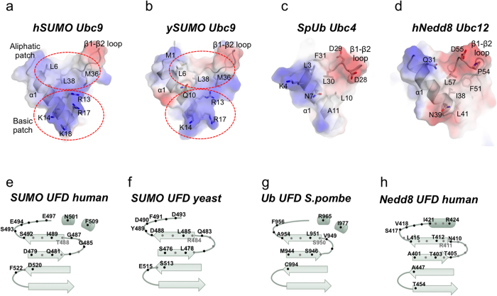
Figure 2. Comparison of the UFD-E2 interface from different UbL systems.
(a) Transparent electrostatic representation of the interface of human SUMO E2 (hSUMO Ubc9) with the UFD domain. Major contacts are labeled and represented in stick configuration. Basic and aliphatic surface patches are indicated by dotted circles. (b) Transparent electrostatic representation of the interface of yeast SUMO E2 (ySUMO Ubc9) with the UFD domain. Major contacts are labeled and represented in stick configuration. Basic and aliphatic surface patches are indicated by dotted circles. (c) Transparent electrostatic representation of the interface of S.pombe ubiquitin E2 (SpUb Ubc4) with the UFD domain. Major contacts are labeled and represented in stick configuration. (d) Transparent electrostatic representation of the interface of human Nedd8 E2 (hNedd8 Ubc12) with the UFD domain. Major contacts are labeled and represented in stick configuration. (e) Schematic representation of the human SUMO E1 UFD domain contacts with the E2 enzyme. (f) Schematic representation of the yeast SUMO E1 UFD domain contacts with the E2 enzyme. (g) Schematic representation of the S.pombe ubiquitin E1 UFD domain contacts with the E2 enzyme. (h) Schematic representation of the human Nedd8 E1 UFD domain contacts with the E2 enzyme. Black and grey spots indicate the orientation of the side chain in the structure regarding the β-sheet plane.

