Figure 1.
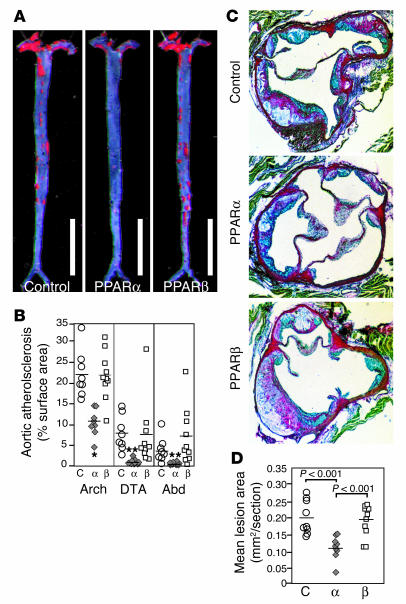
Atherosclerosis in LDLR–/– male mice that were fed the HC diet for 14 weeks. (A) Sudan IV-_stained en face preparations of aortas. Scale bars: 1 cm. (B) Quantitative analysis of atherosclerotic surface area in the entire aorta. (C) Sections through the aortic root at the level of the aortic valves. The micrographs are taken of sections at a similar distance from the aortic root. Original magnification, ×4. (D) Quantitative analysis of lesion areas in the aortic root. Data expressed as the mean ± SEM. C, control; α, PPARα agonist GW7647; β, PPARβ agonist GW0742; Abd, abdominal aorta; Arch, aortic arch. *P < 0.001 and **P _ 0.02, compared with control.

