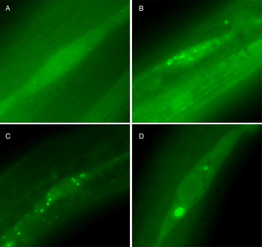
Figure 2.
GFP::LGG-1 expression in hypodermal seam cells of daf-2(e1370) mutants A. daf-2(e1370) mutants grown on OP50 E. coli, at 15°C, display a diffuse localization of GFP::LGG-1. B. daf-2(e1370) mutants grown on OP50 E. coli, at 25°C, display an increase in GFP::LGG-1 positive puncta (up to 12 GFP::LGG-1 positive puncta/seam cell) that represent early autophagic structures or autophagosomes. C. daf-2(e1370) mutants grown on control RNAi E. coli (transformed with empty vector, L4440), at 25°C, display the characteristic GFP::LGG-1 positive punctate structures. D. daf-2(e1370) mutants fed bec-1 RNAi, and raised at 25°C, display an increase in GFP::LGG-1 expression and large GFP::LGG-1 positive aggregates

