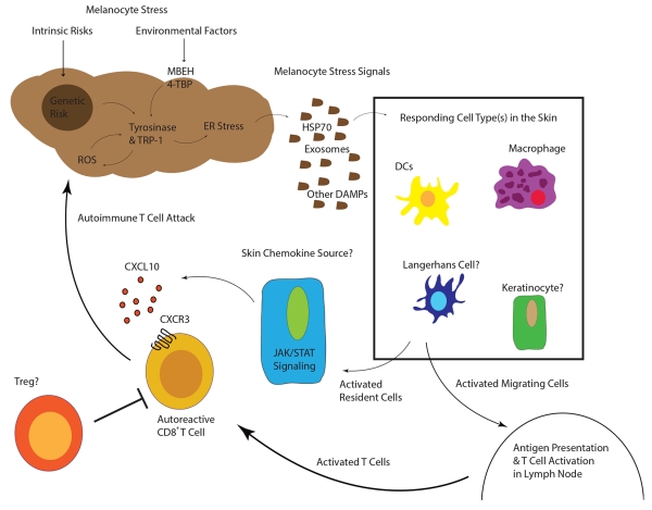
Figure 3.
“Working Model of Vitiligo Pathogenesis”
Inherited genetic risk (HLA (11)(10), XBP1 (16), TYR (13), OCA2 (15), M1CR1 (15)) and environmental insults (MBEH and 4-TBP) induce a state of melanocyte stress, exemplified by ER stress. Stressed melanocytes signal to local innate and resident skin cell types via exosomes containing antigen and DAMPs, soluble HSP70, and/or other factors (30-34). Responding cell types are activated by these signals and some may migrate to the draining lymph nodes where they activate T cells. Other responding cells in the skin secrete chemokines to recruit autoreactive T cells, which are directly responsible for killing melanocytes. In active disease, one or more cell types may respond to IFN-γ and secrete CXCL10 to recruit T cells to the skin where melanocytes reside.

