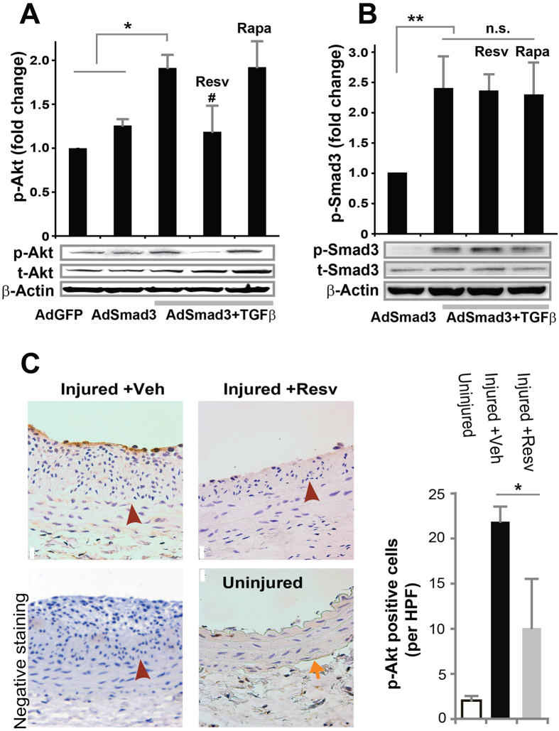
Figure 7. Inhibitory effect of resveratrol on TGFβ/Smad3-stimulated Akt activation in vitro and in vivo in balloon-injured rat carotid arteries.
(A,B) Rat aortic SMCs were infected with AdGFP (control) or AdSmad3 followed by pre-incubation with vehicle (DMSO), resveratrol (50 μM), or rapamycin (200 nM) for 2 h, and then treated with solvent or TGFβ1 (5 ng/mL). For Western blotting analyses of p-Akt (A) and p-Smad3 (B), cells were collected after TGFβ1 treatment for 2 h and 0.5 h, respectively. Quantification: mean ± SEM of three independent experiments; normalization to total Akt (t-Akt) or to total Smad3 (t-Smad3); *P < 0.05 compared to each of the first 2 conditions; **P < 0.05 compared to AdSmad3 only; #P < 0.05 compared to AdSmad3 + TGFβ1 without pre-incubation with a drug; n.s., not significant. (C). Rat carotid arteries were treated with either vehicle or resveratrol and collected on day 14 after angioplasty. Shown on the left are representative sections of p-Akt Immunostaining. Arrows indicate external elastic lamina (EEL); arrowheads mark internal elastic lamina (IEL). Scale bar = 20 μm. Negative staining: IgG instead of a primary antibody was used. Quantification: mean ± SEM of 4–5 animals; *P < 0.05 compared to vehicle control; HPF, high power field.

