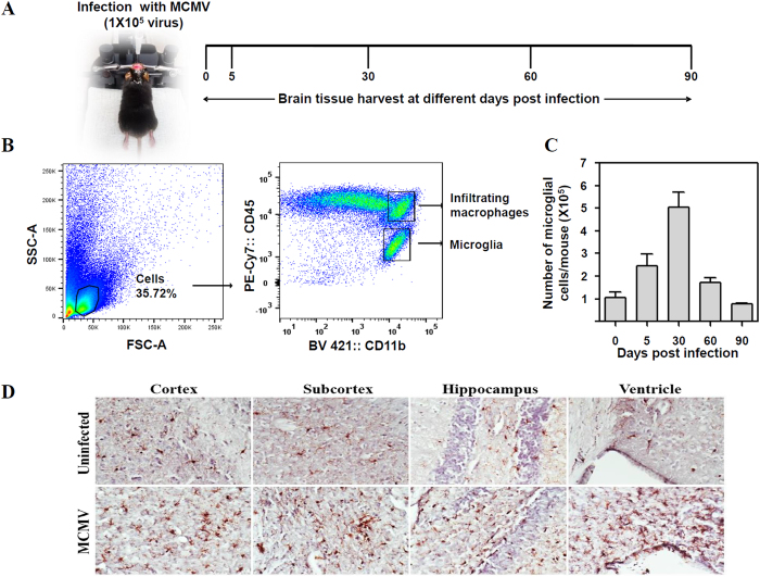
Figure 1. Chronic neuro-inflammation following MCMV-induced encephalitis.
Mice were infected with 1 × 10⁵ TCID50 units (in 10 μl) of MCMV. One group of mice was not infected with MCMV. At 0, 5, 30, 60 and 90 dpi, mice were euthanized and brain tissues were harvested to isolate brain mononuclear cells (BMNCs). (A) Treatment and sampling schedule. (B) The representative flow cytometric dot plots to identify microglial cells. BMNCs were first gated on their forward and side scatter properties followed by gating on CD45 and CD11b. Gating on the CD45intCD11bhi population identified microglial cells. (C) The number of brain resident microglial cells post infection. Counts were acquired on a FACS LSR II H4760 flow-cytometer (using FACS Diva software) and analyzed using FlowJo software (Tree Star, USA). Data presented are mean ± SE of two experiments with 4–6 mice per time point. (D) Immunohistochemical staining of brain sections demonstrating persistence of microglial cells (brown) in the cortex, subcortex, hippocampus and ventricle regions of uninfected and MCMV-infected mice at 30 dpi.

