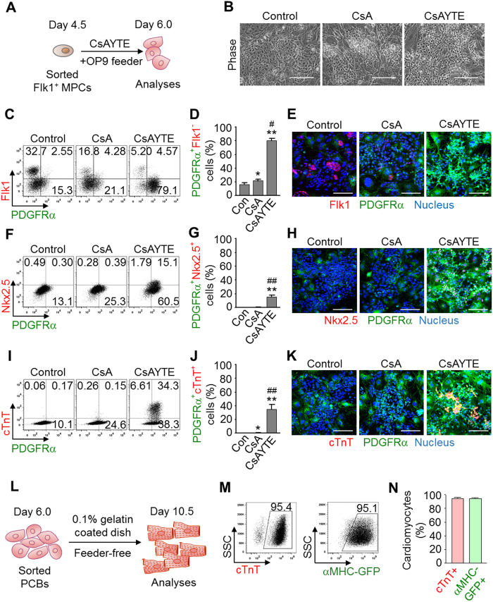
Figure 2. CsAYTE generates PCBs that eventually differentiate into cardiomyocytes.
(A) Protocol to generate PCBs from Flk1+ MPC by CsAYTE stimulation and subsequent analyses. (B) Phase-contrast images showing differentiating Flk1+ MPCs at day 6.0 incubated with control vehicle (Control), CsA, and CsAYTE. Scale bars, 100 μm. (C–K) Representative FACS analyses, quantifications, and images of PDGFRα+Flk1− PCBs, PDGFRα+ Nkx2.5+ cells, and PDGFRα+ cTnT+ cells differentiated from Flk1+ MPCs at day 6.0 incubated with Control, CsA, and CsAYTE (Scale bars, 100 μm). Each group, n = 3–6. *p < 0.05 and **p < 0.01 versus Con; #p < 0.05 and ##p < 0.01 versus CsA. (L) Protocol for analyses of PCB-derived cardiomyocyte differentiation in a feeder-free culture. (M and N) Representative FACS analyses and percentages of PCBs-derived cTnT+ and αMHC-GFP+ cardiomyocytes grown in feeder-free culture. Each group, n = 4–5.

