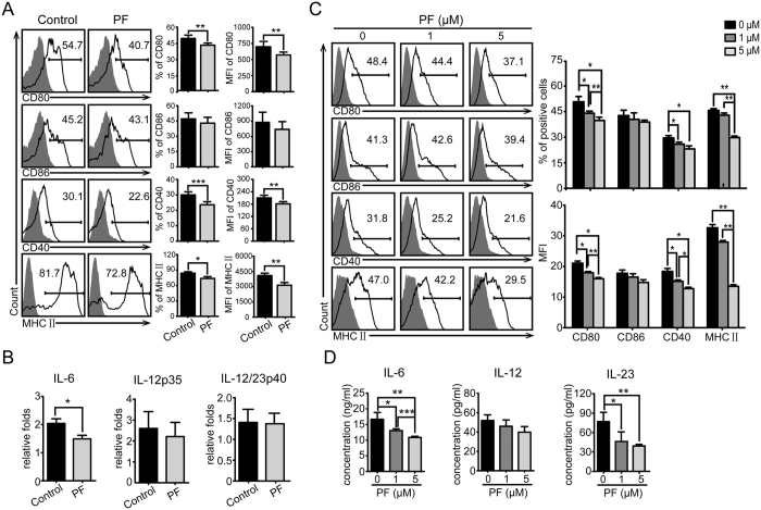
Figure 3. Effect of PF on CD11c+DCs and BMDCs.
(A) Splenocytes from PF-treated or control EAE mice were analyzed for the expression of CD80, CD86, CD40, and MHC II in CD11c+ gate by flow cytometry. (B) mRNA levels of IL-6, IL-12p35, and IL-12/IL-23p40 in CD11c+DC were analyzed by real-time PCR. Data are representative of three independent experiments with eight mice per group. Values were expressed as mean ± SEM. *p < 0.05; **p < 0.01, ***p < 0.001. (C) Bone marrow cells were stimulated by GM-CSF and IL-4 for 5 days to induce BMDCs in different concentrations of PF, and the expression of indicated markers were analyzed by flow cytometry. (D) The production of cytokines in the culture supernatants from BMDCs stimulated with LPS was examined by ELISA. Data are representative of at least three independent experiments. Values were expressed as mean ± SEM. *p < 0.05; **p < 0.01, ***p < 0.001.

