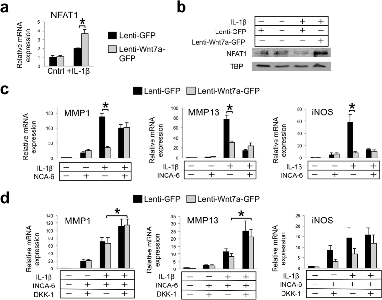
Figure 7. Inhibition of NFAT signaling attenuates the effect of Wnt7a on MMP inhibition in human chondrocytes.
(a) RT-PCR analysis of NFAT1 gene expression in nHACs after infection with lenti-GFP or lenti-Wnt7a-GFP, and cultured with or without IL-1β (5 ng/mL). NFAT1 expression was upregulated with Wnt7a ectopic expression in the IL-1β group only. (b) Western blot analysis of nuclear NFAT1 protein expression after nHACs were infected with lenti-GFP or lenti-Wnt7a-GFP and cultured with or without IL-1β (5 ng/mL). TBP served as a loading control. Original films for cropped images can be found in the supplementary information file. (c) RT-PCR analysis of MMP1, MMP13 and iNOS after treatment with 20 μM of INCA-6 on nHACs infected with lenti-GFP or lenti-Wnt7a-GFP and treated with or without 5 ng/mL IL-1β for 2 days. (d) RT-PCR analysis of MMP1, MMP13 and iNOS gene expression after treatment with 20 μM of INCA-6 and/or 250 ng/mL DKK-1 on nHACs infected with lenti-GFP or lenti-Wnt7a-GFP and cultured in the presence of absence of IL-1β (5 ng/mL). Each experiment had three biological repeats/treatment, and at least three independent experiments were performed. Analysis of variance (ANOVA) with post-hoc tests was used for evaluating the statistical significance between the gene expression of lenti-GFP and lenti-Wnt7a cells across all of experimental conditions. All data are shown as mean ± SEM. *p < 0.05.

