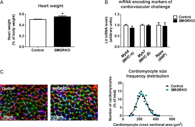
Figure 4.
Heart weight is elevated in 6-week-old male SMGRKO mice, but there is no evidence of pathological cardiomyocyte hypertrophy. (A) Heart weight is elevated in 6-week-old male SMGRKO mice (black bar) compared with littermate controls (white bar) (N = 10–11). (B) Levels of mRNA encoding markers indicative of pathological cardiac remodelling, including myosin heavy chain alpha (Myh6), myosin heavy chain beta (Myh7) and atrial natriuretic peptide (Nppa), were not altered in LV of 6-week-old male SMGRKO mice compared with controls (N = 10–11). (C) Representative images of formalin-fixed, paraffin-embedded sections of LV of 6-week-old male SMGRKO mice and controls showing wheat germ agglutinin staining of plasma membrane (red), isolectin B4 staining of the vasculature (green) and DAPI staining of nuclei (blue). The frequency distribution for cardiomyocyte cross-sectional area did not differ in SMGRKO mice (blue line) compared with controls (black line). N = 10; 40 cardiomyocytes measured/heart. Data are means ± s.e.m. and were analysed using an unpaired t-test, *P < 0.05.

 This work is licensed under a