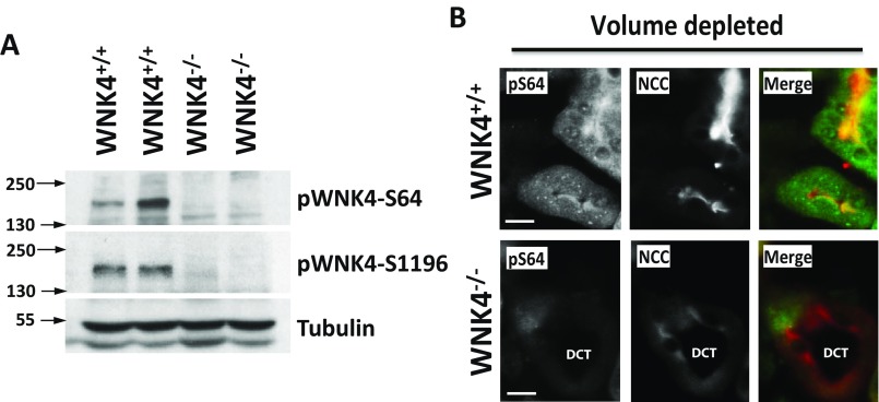
Fig. S5.
Demonstration of specificity of pWNK4 antibodies for in vivo studies. (A) Kidney lysates from WNK+/+ and WNK4−/− mice were blotted with the different WNK4 phosphoantibodies. Only the pS64 antibody and the previously generated pS1196 antibody (50) gave a specific signal. This is, a band of the expected size was observed that was absent in the samples from the WNK4−/− mice. Nonspecific binding with the remaining four antibodies, masked the bands corresponding to phospho-WNK4. (B) Kidney sections from volume depleted WNK4+/+ and WNK4−/− were stained in parallel with pWNK4 antibodies. Only the pS64 antibody gave a specific signal that was not observed in the WNK4−/− mice. This signal was observed in NCC-positive tubules (DCTs). Note that a lower NCC signal was observed in the WNK4−/− mice. This was expected as it has been previously reported that NCC expression is greatly decreased in WNK4−/− mice (5). (Scale bars, 10 μm.)

