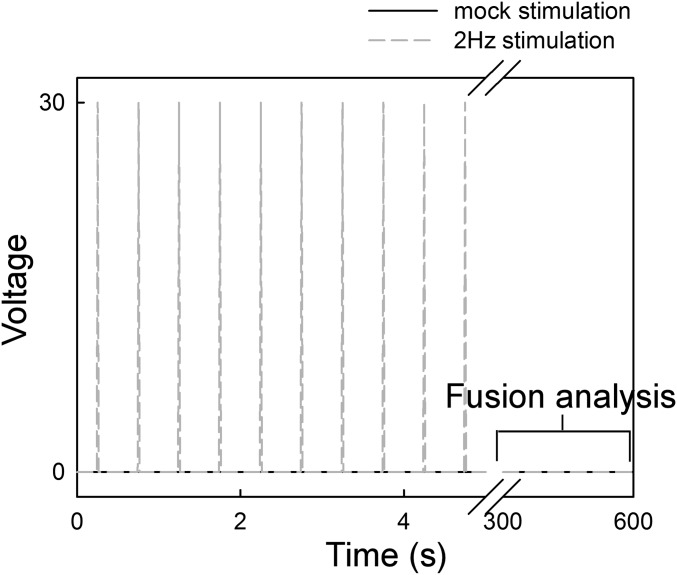
Fig. S4.
Pacing scheme applied in Fig. 5D. Freshly isolated in vivo-transformed ventricular myocytes plated on top of laminin-coated coverslips were mounted in a custom-made chamber equipped with platinum electrodes. At the confocal microscope stage, the chamber was connected to a Grass stimulator, and 2-Hz square-shaped pulses were triggered (30 V, 2 ms) for 5 min. The stimulation buffer was 0.25% BSA CaCl2 without BDM. After brief and careful washing, and loading of the same experimental chamber with 10 mM BDM-supplemented 0.25% BSA ECM, fusion events were evaluated for 5 min. Mock experiments involved the same treatment of the cells without field stimulation.

