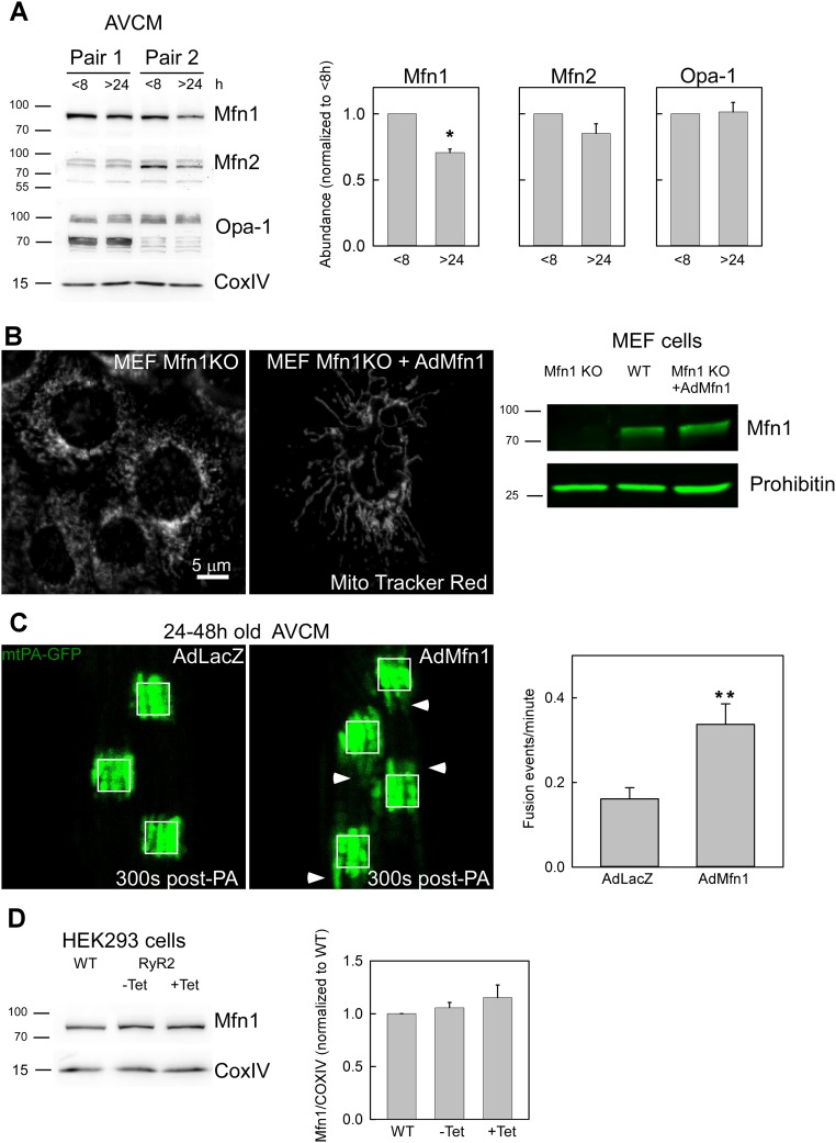
Fig. S5.
Mitochondrial fusion protein Mfn1 decays in cultured AVCMs and rescues fusion upon exogenous expression. (A) Freshly isolated AVCMs were plated on culture dishes for 2–8 h or kept in culture for at least 24–30 h. Samples were snap-frozen, and membrane extract was prepared for Western blot analysis and tested with the indicated antibodies. Specific bands were quantified with regard to the <8-h condition. n = 4 independent experiments. P < 0.05. (B) Mfn1 KO cells were transduced with AdMfn1, and at 24–28 h after infection, the cells were loaded with MitoTracker Red and evaluated by confocal microscopy (Left). Cell membrane extracts were evaluated by Western blot analysis to determine the abundance of Mfn1 in MEF WT, Mfn1 KO, and Mfn1 KO cells transduced with AdMfn1 (Right). Transformation of Mfn1 KO cells with AdMfn1 rescued the expression of Mfn1. (C) AdmtPA-GFP and AdmtDsRed in vivo-infected AVCMs were infected in vitro with either control AdLacZ or AdMfn1 virus and then evaluated at 24–48 h after plating. The white squares represent 25-µm2 regions. (Left) Representative images of mtPA-GFP spreading at 300 s post-PA shows increased mitochondrial continuity for AdMfn1 transformed cells compared with control, as indicated by white arrowheads. (Right) Quantification of mitochondrial fusion for the LacZ- and Mfn1-infected AVCMs shows an increase in mitochondrial fusion for Mfn1 cells (n = 39) compared with LacZ cells (n = 40). **P < 0.01. (D) WT and RyR2-HEK293T cells noninduced or induced for 48 h with tetracycline (Tet) were used to prepare membrane-enriched extracts, which were evaluated on 8% PAGE and tested for Mfn1 protein levels. n = 2 independent experiments.

