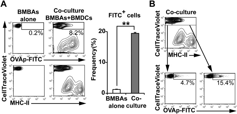
Fig. S5.
FITC-labeled OVA peptides are transferred from DCs to basophils. BMBAs were cultured for 12 h with or without BMDCs that had been pulsed with FITC-conjugated OVA peptides (OVAp-FITC) for 8 h. (A) Staining profiles of CellTrace Violet, FITC and MHC-II are shown (Left). (Right) MFI of FITC on cultured BMBAs is summarized (mean± SEM; n = 3 each). (B) (Upper) Staining profile of CellTrace Violet and MHC-II. (Left) Staining profiles CellTrace Violet and FITC in MHC-IIlo CellTrace Violethi cells and MHC-IIhi CellTrace Violethi cells. Data in A and B are representative of at least three independent experiments.

