Fig. S2.
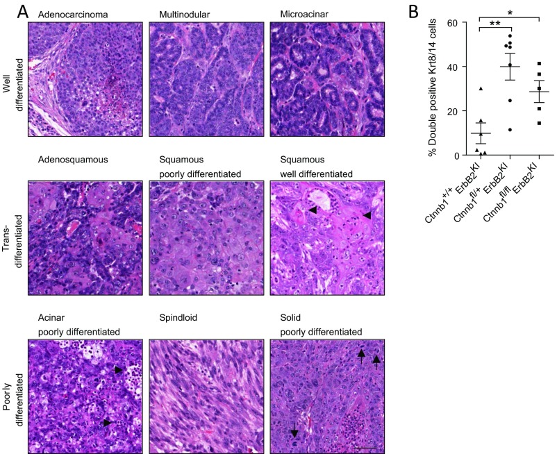
Loss of β-catenin promotes a diverse spectrum of tumor pathologies, including features of poor differentiation and an expansion of a bipotent progenitor population. (A) End-point ErbB2KI tumors, regardless of β-catenin KO status, were subjected to H&E staining to illustrate various pathological subtypes. Tumor pathologies are classified into three main categories: well-differentiated, transdifferentiated, and poorly differentiated. Well-differentiated tumors still retain features of normal epithelial cell organization, such as cell to cell contact and partially intact basement membrane. Microacinar-type tumors contain tumor units resembling normal ducts. Transdifferentiated tumors contain cells with squamous morphology and often abundant keratin deposits (arrowheads). Poorly differentiated acinar tumors exhibit abundant leukocyte infiltration (horizontal arrows) as well as tumor cells that fail to arrange in organized structures, such as those observed in well-differentiated acinar type. Less common forms of poorly differentiated tumors include spindloid (EMT-like) tumors and solid tumors with leukocyte infiltration, pleomorphic nuclei, and abundant mitotic figures (vertical arrows). (Scale bar: 40 μm.) (B) End-point tumors were costained for Krt8 and Krt14 by IHF. Confocal images of 10 fields within the tumors were taken, and double-positive Krt8/14 cells were scored and presented as percentages of the total number of cells. Error bar: SEM. *P < 0.05 (Student’s t test); **P < 0.005 (Student’s t test).

