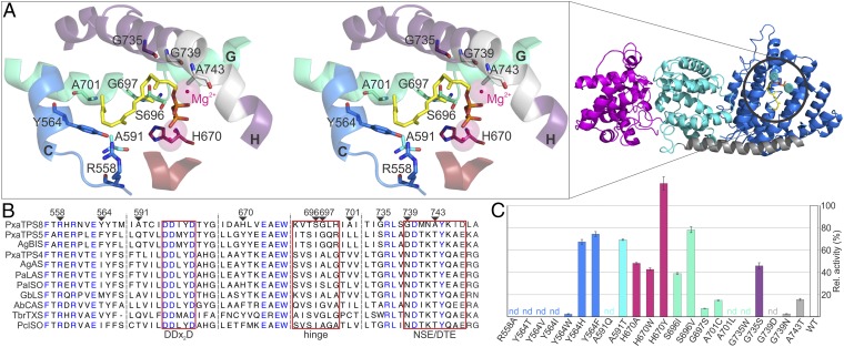
Fig. 4.
(A, Right) Homology model of PxaTPS8 based on the structure of A. grandis α-BIS (PDB-ID: 3EAS) (19) showing the typical diTPS three-domain structure including the γ-, β-, and α-domains (magenta, cyan, and blue, respectively). (Left) Stereoview of the PxaTPS8 active site with docked GGPP (yellow), DDxxD motif (red), and NSE/DTE motif (gray). Analyzed residues and helices H and G (containing the nonhelical hinge region) are color coded. (B) Alignment of select PxaTPS8 amino acids with known gymnosperm TPSs (for abbreviations Table S1). (C) Pseudolaratriene formation by PxaTPS8 variants relative to the wild-type enzyme quantified with eicosene as an internal standard. Color coding is as in A.

