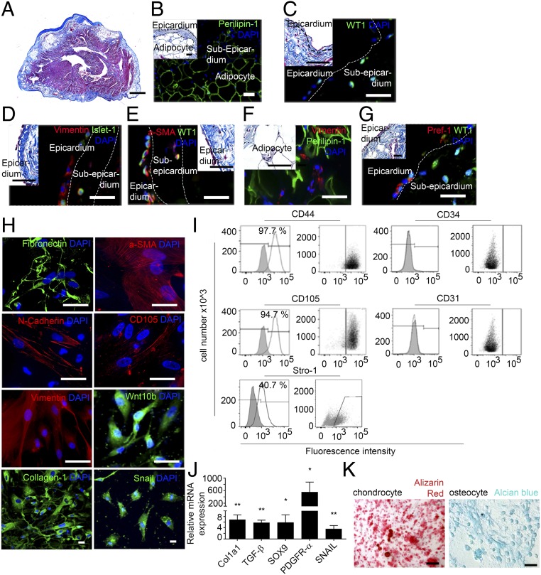
Fig. 1.
Human atrial epicardial progenitor cells have mesenchymal properties. (A) Masson’s trichrome staining of 7-µm-thick sections of human atrial tissue (n = 26). (Scale bar, 100 µm.) (B–G) Immunofluorescence staining in human atrial tissue sections (n = 8) for Perilipin-1 (B), WT1 (C), vimentin and Islet-1 (D), α-SMA and WT1 (E), vimentin and Perilipin-1 (F), and Pref-1 and WT1 (G). (Scale bars, 20 µm.) Insets show Masson’s trichrome staining. (Scale bars, 10 µm.) (H) Immunofluorescence staining of aEPDCs for fibronectin, α-SMA, N-cadherin, CD105, vimentin, Wnt10b, collagen-1, Snail, and DAPI (n = 10). (Scale bars, 10 µm.) (I) Flow cytometry of aEPDCs for CD44, CD105, CD31, CD34, and Stro-1 markers (n = 10). Specific isotype controls are shown in gray. (J) Quantitative PCR (qPCR) analysis for Col1a1, TGFβ, SOX9, PDGFRα, and SNAIL in aEPDCs. Data are expressed as the fold change relative to unpassaged aEPDCs (n = 15) and represent the mean ± SEM of independent experiments. *P < 0.05, **P < 0.01, one-way ANOVA and Bonferroni’s post hoc test. (K) Bright-field images of aEPDC-derived chondrocytes or osteocytes revealed by alizarin red or Alcian blue staining, respectively (n = 3). (Scale bars, 20 µm.)

